Showing posts with label aging. Show all posts

Monday, July 23, 2018

Modeling the Telomere length distribution with Chromosomal Segregation along Synchronous Cellular Divisions

Telomeres: structure and functions

If it is necessary to explain what the telomeres are, let's watch some lectures:
First from Elisabeth Blackburn:


Or from Titia de Lange:
 

Modeling Telomere Dynamic:

Levy et al. in Telomere End-replication Problem and Cell Aging, modeled the distribution of telomeric erosion with cells divisions at a single telomere. The model accounts for the decrease in somatic cell division capabilities with in-vitro passages of fibroblasts (Hayflick's limit).

Here, the telomere length of several chromosomes is modeled numerically with segregation of chromosomes in daughter cells. The model takes the 5' degradation of the CCCTAA strand into account. The aim of the simulation is to compare the distributions with QFISH data. The model shows that the telomeres length at pter and qter are correlated for a given homologous chromosome. It can be used to derive statistical tests to detect telomere length difference between homologous chromosomes.

Code Installation

Download several python modules and the jupyter notebook in the same directory:
  • WatsonCrick.py
  • ClassChromosome.py
  • Genome.py
  • Cellular.py
  • telomere-length-distribution-in-synchronous-dividing-cells-ipynb
Some unit tests can be run, a jupyter notebook is available (see the link to gist), they yield:

Model structure:

 The  model consists in a class Cell which have a Genome, which have chromosomes, which have two complementary single strands DNA (Watson/Crick). Each strand has two telomeres.

A single strand object can be instantiated as follow:


A chromosome object is instantiated with two complementary Watson/Crick strands:


For example, two chromosomes, rosalind and franklin can be instantiated as follow:

A single chromatid chromosome, in the G1 state of the cell cycle, can be triggered to the G2 state:
After a round of DNA replication, 5' CCCTAA motifs are incompletely replicated and TTAGGG 3' can be randomly degraded, leading to shorter telomeres on metaphasic chromosomes on both chromatids at pter and qter. Iin the G2 state a chromosome has two chromatids, the length of a telomere on a given arm (pter ou qter) is given by two values:
The mitosis is triggered by chromosome segregation:
The ros chromosome object keeps its signature before and after the mitosis:
Signature of the ros chromosome in the G2 state before mitosis

Definition of a Genome object:

It is convenient to define a genome object, it is instantiated for example with two chromosomes (2N=2):


The genome state (G1/G2), the chromosomes of the genome can be accessed to read the telomere length (CCCTAA or TTAGGG motifs), the length difference between homologous telomeres:

Cell object has a genome:

Unit test results for Cell object

Once a single genome object is instantiated, a single cell object can be instantiated. Let's build a cell with 2N=10:

The length of the telomeres (bp) in that single cell can be read from a pandas dataframe:

Let's allow 10 synchronous cellular divisions from 1 cell to 1024 cells. Then as for fibroblasts cultures, let's make passages, that is one allows only half randomly chosen cells to divide once:

Results

The envelope of the telomere length distribution at a single telomere after 14 divisions (10 divisions + 4 passages) seems to be gaussian:

Correlation between the length at pter and qter on the same homolog

The length of the telomeres (pter, qter) of the same homologous chromosome (the maternal one for example) are correlated:
It is possible to plot the telomeres length of two homologous chromosomes:

The length of the telomeres belonging to different homologous are not correlated.

Let's plot the length of the telomere from the paternal chromosome 1 at qter as a function of the length of the telomere at 1qter on the maternal homolog:

Mean telomere length in synchronous dividing cells population:

In telomerase negative cells as modeled here, the telomere length decreases with cells divisions.
The decrease of telomere length depends on the 5' exonuclease activity. The amount of degraded DNA is modeled here by random variable, X, following a binomial law (N=400, p=0.4) :

Distribution of X: P(130<X<200)=0.999
to fit the data of Makarov et al. (1997) as follow:


 The expected value of X, the deletion length, is :

taking the RNA primer deletion (20 bp), the mean length of the 3' s G strand is:

160 +20 bp

As published in 1992, the standard deviation of the telomere length increase at each cell division.
So the mean telomere length, mean+/std, was plotted as a function of the cell divisions after an expansion of ten cellular divisions and four passages.

The mean telomere length decreases by 90 bp/div

and the heterogeneity increases:
At each passage the length of each telomere (column) of each cell (row) of the simulation is copied in a pandas data-frame, for example at passage P4:


 Then the mean telomere length in the whole cells population can be calculated:


Using seaborn, the telomere length of some homologs can be compared:

Population of Mixed Cells

Cells from different passages can be mixed, for example in equal proportions. The correlation between pter and qter of a given homolog should start to vanish:

Senescence in-silico:

Short telomeres trigger an irreversible transition to G0 state of the cell cycle (senescence). Let's take a cells population, make successive passages. Initial cells population is expended from one cell (telomerase off) after eight  divisions. The genome of the initial cell is instantiated with 2N=4 and with the length of the shortest telomere set to 2000 bp.


At each passage, it's possible to count the cells in the G0 state or to calculate the confluence of the cells population. The threshold telomere length triggering the G0 state is set arbitrarily to 200 bp.  The initial population is passed 20 times:

The confluence is calculated as the number of cells at a given passage by the initial number of cells. With the shortest initial telomere length set to 2000 bp, the confluence reach 50% after 18 doublings:


Senescence in silico: confluence  (100 N cell/ N initial cells) decreases with cells divisions

Python modules and jupyter notebook


Monday, March 19, 2012

Genomic head to head telomeric sequences detection by one primer PCR

Introduction
Dicentric chromosomes are the signature of recombination events which may imply telomeric motifs. To observe dicentric chromosomes in senescent cells, a cell culture  followed by an enrichment step is performed to get enough metaphasic cells. However metaphasic cells become difficult  to get as the cell culture reaches senescence or if cells differenciate, thus most of  fusions between short telomeres, when they occur, remain hidden in interphasic cells.
Telomeric fusions can generate head to head TTAGGG motifs as:
(TTAGGG)n(CCCTAA)m

Head to head telomeric sequences (blue) at a fusion point (several possibilities), flanked by proximal (orange, green) and distal (purple) subtelomeric sequences.


 Such kind of sequence is known in the human genome at the 2q13-q14 locus (Ijdo et al.).

>gi|180516|gb|M73018.1|HUMCHR2A Human ancestral telomeric fusion DNA sequence
GTGCCCCGGCGCCACGAGGGCGCTGGCGACCACTGTAAGCAAGAGAGCCCTGCGCCTCTCTGCGCCTGCG
CGGCCGGGCCGGCCGGCCGCCTTTGCGATGGCGGAGTTGCGTTCTCCTCAGCACAGACCCGGAGAGCACC
GCAGGGCGACCTGCGTTGTCTCTGCACAGATTTCGGTGGTACTGCGAAGGCGGACGAGAGTTCTCCTCAG
GTCAGACCCGGGCCGGGCCGGCTAGGATACCGCGAGGCGAGCTGCGTTCTGCTCAGCACAGACCTGGGGG
TCACCGTAAAGATGGAGCAGCATTCCCCTAAGCACAGAGGTTGGGGCCACTGCCTGGCTTTGTGACAACT
CGGGGCGCATCAACGGTGAATAAAATCTTTCCCGGTTGCAGCCGTGAATAATCAAGGTCAGAGACCAGTT
AGAGCGGTTCAGTGCGGAAAACGGGAAAGAAAAAGCCCCTCTGAATCCTGGGCAGCGAGATTATCCCAAA
GCAAGGCGAGGGGCTGCATTGCAGGGTGAGGGTGAGGGTGAGGGTGAGGGTTAGGGTTTGGGTTGGGGTT
GGGGTTGGGGTTGGGGTAGGGTTGGGGTTTGGGTTGGGGTTAGGGTTAGGGGTAGGGGTAGGGTCAGGGT
CAGGGTCAGGGTTAGGGTTTTAGGGTTAGGGTTAGGGTTAAGGTTTGGGGTTGGGGTTGGGGTTGGGGTT
AGGGGTTAGGGGTTAGGGGTTAGGGTTGGGGTTGGGGGTTGGGGTTGGGGTTAGGGGTAGGGGTAGGGGT
AGGGTTAGGGTTAGGGTTAGGGTAAGGGTTAAGGGTTGGGGTTGGGGTTGGGGTTAGGGTTAGGGGTTAG
GGTTAGCTAACCCTAACCCTAACCCCTAACCCCTAACCCCAACCCAAACCCCAACCCCAACCCCAACCCT
ACCCCTACCCCTAACCCCAACCCTTAACCCTTAACCCTTAACCCTTACCCTAACCCTAACCCAAACCCTA
ACCCTAACCCTACCCTAACCCAACCCTAACCCTAACCCTACCCTAAGCCTAAAACCCTAAAACCGTGACC
CTGACCTTGACCCTGACCCTTAACCCTTAACCCTTAACCCTAACCCTAACCATAACCCTAAACCCTAACC
CTAAACCCTAACCCTACCCTAACCCCAACCCCTAACCCTAACCCCTATACCCTAACCCTAACCCTACCCC
TACCCCTAACCCCAACCCCAGCCCCAACCCCAACCCTTACCCTAACCCTACCTAACCCTTAACCCTAACC
CCTAACCCTAACCCCTAACCCTACCCCAACCCCAAACCCAACCCTAACCCAACCCTAACCCAACCCTAAC
CCCTACCCTAACCCCTAACCCTAACCCCTACCCTAACCCCTAACCCTAACCCCTACCCTAACCCCTAACC
CTAGCCCTAGCCCTAACCCTAACCCTCACCCTAACCCTCACCCTAACCCTCACCCTCACCCTCACCCTCA
CCCTAACCCAACGTCTGTGCTGAGAAGAATGCTCGTCCGCCTTTAAGGTGCCCCCCAGGTCTGTGCTGAA
CAGAACGCACGTCCGCCGTCGCAGTGCCCTCAGCCCGGGTCTGACCTGAGAAGAACTCTGCTCCGCCTTC
GCAATAGCCCCGAAGTCTGTGCAGAGGAGAACGCAGCTCCGCCCTCGCGATGCTCTTCGGCTGTGTGCTA
AAGAGAACGCAACTCCGCCCTCGCAAAGGCGGCGCCGCCGCGGAGGCCGGAGAGGCGCGGCGCCGCGGAG
GCCGGAGAGGCGCGGCGCGCGGGAGGCCGGAGAGGCGCGGCGCCGCGGAGGCCGGAGAGGCGCGGCGCCG
CGGAGGCCGGAGAGGCGCGGCGCCGCGGAGGCCGGAGAGGCGCGGCGCCGCGG

The fusion point of this sequence is:
..TTAGGGTTAGCTAACCCTAA..
It is the cicatrix of an ancestral chromosomic fusion occured during karyotype evolution. As the human 2q13-q14 contains a short amount of degenerated telomeric motifs these motifs are degenerated so they cannot be detected by QFISH with (CCCTAA)3 PNA probe (most of the time):
Possible QFISH detection of telomeric motifs at 2q13-q14 on one HSA2 homolog. Interstitial telomeric motifs are detected at the junction of euchromatin/heterochromatin on the Y chromosome. (Red: telomeric (CCCTAA)3-PNA probe, contrast was increased after background substraction)


Using Polymerase Chain Reaction and only one (TTAGGG)n primer, would it be is possible to detect head-to-head genomic telomeric sequences possibly resulting from chromosomal fusion ?

Detection of a genomic head-to-head telomeric sequence by PCR amplification with one (TTAGGG)n primer (Dark Blue big arrow)
Here was investigated the possibility to detect somatic telomeric fusion occurring in senescent or SV40-transformed fibroblasts by one primer PCR amplification of head-to-head telomeric sequences.(J-P Pommier, unpublished data)

It was found that PCR products ( next called G PCR products) can be produced from genomic human DNA with single TTAGGG primers. It is known that human genome DNA contains head-to-head telomeric motifs and possibly some were generated by chromosomal instability in cultured cells.

Material and method:
The  cycle conditions of the first PCR assay were inspired by the paper of Ijdo et al. . As no PCR product was detected with these conditions, the stringency conditions were relaxed by decreasing the annealing temperature, without my lab-book, I cannot bring more precision on the cycling conditions.
Two kinds of assay were performed: G-PCR with (TTAGGG)n primers (n=3 or 5) or C-PCR with (CCCTAA)n primers (n=3 or 5) , both with 100ng of genomic DNA (high molecular weight placenta DNA, primary fibroblasts, SV40 transformed fibroblast pre-crisis/post-crisis, wild type S. cerevisiae genomic DNA was also tested). In a second assay, both G an C primers were used at high (100 nM?) or low (20 nM?) concentration. G3-primer and C3-primer had 5' non telomeric sequence allowing a second PCR round with a non telomeric primer.
In an assay, prior PCR, 1 microgramme of genomic  DNA was digested by Hpa II (methylation sensitive) or Msp I restriction endonuclease (NEB) targeting GGCC or by an other endonuclease targeting a TTAA restriction site. A 100ng aliquot was then submitted to G-PCR.
PCR products were analysed in a 2-3% agarose gel electrophoresis, stained with ethidium bromide, with a molecular weight standard in the range 200 to 2000 bp approximatly (possibly molecular weight marker IV, Roche?).

Results:
Genomic DNA from placenta, primary fibroblasts, precrisis SV40 transformed fibroblasts submitted to G5-PCR (wells n°6,8,10) yield a smear like PCR product with possibly discrete bands. The same genomic DNA submitted to C5-PCR yield a smears as PCR product with no bands.
From left to right: Wells n°2, 13: molecular weight markers. Wells n°6, 8, 10: G5-PCR with (TTAGGG)5 primer. Wells n°7, 9, 11: C5-PCR with (CCCTAA)5 primer. Well n°12:?. The wells n°3, 4 may be some control (no primer, no genomic DNA).

More conditions were tested in the following assay:
C5-PCR or C3-PCR yield a smear as PCR product at high concentration. The PCR product becomes undetectable at low primer concentration. I performed C5-PCR or C3-PCR as a control, I didn't expect to detect a PCR product with a (CCCTAA)n primer due to the head to head orientation of the telomeric motifs in a telomeric fusion. Queue-to-queue telomeric motifs may occur, but those PCR products are likely PCR artifacts.
More interestingly, G5-PCR and G3-PCR yield bands-like PCR products with human DNA in all conditions:
  • Bands are observed with human DNA as PCR target, whereas a smear is observed with yeast DNA, indicating that the PCR product detect a specific structure in the human genome which is not present in the yeast genome.
  • With the "eyes of faith" (les yeux de la foi), G5-PCR at hight concentration primer, seems to distinguish postcrisis DNA (well 8, upper) from normal or precrisis DNA (wells 6,7 upper) by an relative increase of the amount of the short band. As the SV40 transformed fibroblasts reach the crisis, the frequency of dicentric chromosomes increase and become stable when an immortal clone emerge from the crisis. It is consistent with an accumulation of head-to-head telomeric motif generated by chromosomal fusion between short telomeres.
  • The shortest band (~600 bp) vanishes from the PCR product as the G5 primer concentration is diminished regardless the source of DNA (primary cells, precrisis or postcrisis cells). There are possibly several ancestral HTH telomeric motifs per cell, whereas there is less than one neo HTH telomeric motif per cell (statistically around the crisis, cells have 0 or 1 dicentric chromosomes, even some cells have more). As neo HTH telomeric motifs are rare events, decreasing G5 primer would decrease PCR detection efficiency. The same for G3 primer due to thermal stability.
In the last assay the presence of palindromic sequences in potential HTH telomeric motifs was tested with G5-PCR only.
Head-to-head telomeric motif generation by chromosomic fusion

Some of them generate palindromic sequences as GGCC cut by the isoschisomeres Hpa II / Msp I restriction endonucleases, or TTAA .There are 36 possible head-to-head telomeric motifs as follow:


TTAGGG
TAGGGT
AGGGTT
GGGTTA
GGTTAG
GTTAGG
CCCTAA
TTAGGGCCCTAA
TAGGGTCCCTAA
AGGGTTCCCTAA
GGGTTACCCTAA
GGTTAGCCCTAA
GTTAGGCCCTAA
CCTAAC
TTAGGGCCTAAC
TAGGGTCCTAAC
AGGGTTCCTAAC
GGGTTACCTAAC
GGTTAGCCTAAC
GTTAGGCCTAAC
CTAACC
TTAGGGCTAACC
TAGGGTCTAACC
AGGGTTCTAACC
GGGTTACTAACC
GGTTAGCTAACC
GTTAGGCTAACC
TAACCC
TTAGGGTAACCC
TAGGGTTAACCC
AGGGTTTAACCC
GGGTTATAACCC
GGTTAGTAACCC
GTTAGGTAACCC
AACCCT
TTAGGGAACCCT
TAGGGTAACCCT
AGGGTTAACCCT
GGGTTAAACCCT
GGTTAGAACCCT
GTTAGGAACCCT
ACCCTA
TTAGGGACCCTA
TAGGGTACCCTA
AGGGTTACCCTA
GGGTTAACCCTA
GGTTAGACCCTA
GTTAGGACCCTA

If the telomeric fusion point contains a palindromic sequence, submitting the genomic DNA to endonuclease prior G5-PCR may prevent the PCR amplification:
Endonuclease digestion of DNA may prevent G-PCR amplification.
In uncut DNA (primary fibroblasts (line 1), precrisis fibroblasts (line 6), immortalized clone (line 11)), G5-PCR produces two major bands bellow 1200 bp. Those bands vanished as DNA is cut with MspI (GGCC) or with an endonuclease targeting TTAA sequence (from left to right, primary cells, pre or post-crisis fibroblasts:lines 2, 4, 7,9, 12, 13), indicating that palindromic sequences exist between inverse tandem telomeric motifs.
When genomic DNA is cut with HpaII, an endonuclease cutting GGCC motifs free of methyl-cytosine, the G-PCR product cannot be distinguished from the uncut DNA (lines 3, 8, 13), indicating that the cytosines are methylated in/between inverse telomeric motifs.

G5-PCR following endonuclease cut (Agarose gel + EtBr).(well 1: primary fibroblast DNA (PF) uncut, well2: PF+MspI, well3: PF+HpaII, well4: PF+TTAA cut, well5: molecular weight markers (possibly :2176, 1766, 1230, 1033, 653, 517, 453 bp), well6: precrisis transformed (PT) fibroblast uncut, well7: PT+MspI,well8: PT+HapII, well9: PT+TTAA cut, well10: mw marker(?), well11: Immortalized postcrisis fibroblast (IF) uncut, well12: IF+MspI, well13: IF+HpaII, well14: IF+TTAA cut.

Discussion:
If the aim  was to detect somatic telomeric fusions associated to cellular senescence or to cellular transformation, the G5-PCR assays failed to distinguish clearly the different cell states. The G-PCR to detect end-to-end telomeric fusion may be optimized (PCR optimization) by increasing stringency:
  • by increasing annealing T°
  • by decreasing elongation time
  • using hot-start PCR (Taq hot-start or wax)
Prior PCR, genomic DNA could be digested by Bal31 to completely remove terminal telomeric motifs and to keep telomeric motifs belonging to potential fusion points.
Polyacrylamide gel electrophoresis (PAGE) with P32-labelled (TTAGGG)n primer  instead of ethidium-bromide stained agarose gel to visualize PCR product, may detect somatic telomeric fusions in the range 0-600 bp. In the last example, the six last lines correspond to G-PCR products were visualized by PAGE (may be products corresponding to lines 6 to 11 in the second assay).
Real time PCR, in-situ PCR on nuclei could be also considered.
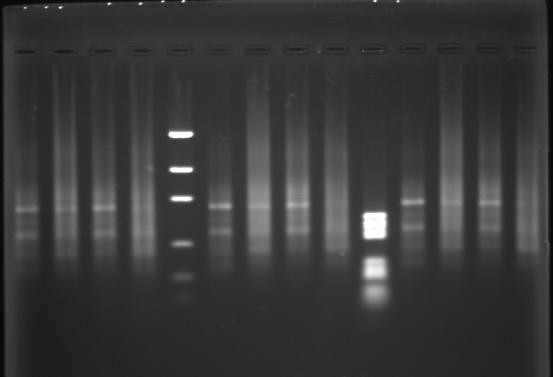
Six G5-PCR products (right side) in a PAGE+Ethidium Bromide staining
I was expecting that detection of somatic telomeric fusions yields short PCR products, bellow 500 bp. The bands in the 1Kb range, are not so short. They may correspond to head-to-head telomeric motifs flanking non telomeric sequences (containing TTAA and methyl-C GGCC motifs).
Two kinds of sequences leading to bands in G-PCR product.
This last hypothesis could be checked by FISH. The G-PCR product could be labelled (after a second round of PCR) with Fluo-dNTP and hybridized on metaphasic chromosomes. Three FISH conditions could be set:
  1. labelled G-PCR product alone: some locci should be detected.
  2. labelled G-PCR product+excess of Cot1 DNA: should compete satellite DNA. Detected locci should contain non satellite sequence, possibly telomeres or unique sequences.
  3. labelled G-PCR product+excess of unlabeled telomeric DNA (produced by PCR, Ijdo & al.): detected locci should not contain telomeric motifs.
Of course, G-PCR product should be insensitive to Bal-31 digestion of the genomic DNA prior PCR amplification.